
New science breakthrough is discovering even more about our cells
What’s inside our cells? We learn in school about the nucleus and mitochondria, but scientists today know there’s much more going on in our bodies at a microscopic level. Artificial intelligence is helping researchers to discover even more about what’s going on at a cellular level. The results have proven surprising.
How cellular health influences longevity

Many diseases we suffer from as we age can trace their roots to things going wrong in our cells. DNA mutations, caused by UV light exposure and oxidative stress, can lead to cell death and cancer. Mitochondrial dysfunction, often a result of metabolic processes, can cause degenerative diseases. Cell disorders of all kinds are responsible for diabetes, heart disease, Parkinson’s, Alzheimer’s, organ failure, and more.
Researchers have investigated the role that cell health plays in longevity. They hope to understand how these diseases start, and what we can do to prevent them. But to get a full picture of what’s going on at a cellular level, we first need to know what exactly is inside our cells.
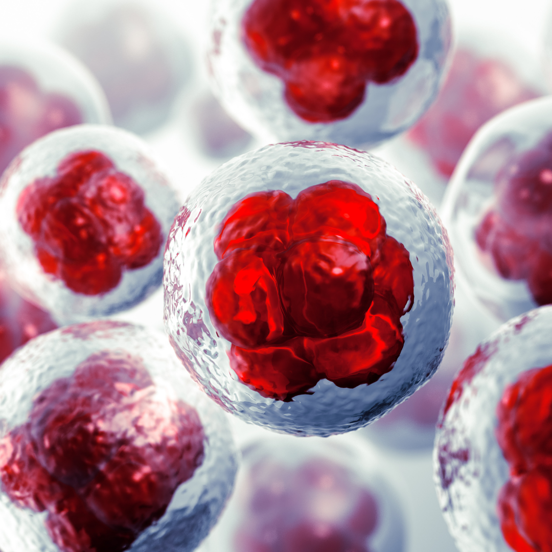
How to see inside our cells

The problem with discovering what’s happening inside cells is they are very small. Even the best microscopes can only see so much. The most powerful microscopes, called scanning tunneling microscopes, can see atoms, which are about 1/10th of a nanometer in size. They don’t work by light like regular microscopes, but use electrical currents to detect movement at an atomic level. They can only work on certain surfaces, and in ultra-high vacuum environments at absolute zero (−459.67 °F).
Light microscopes, like you’d find in a science lab, can see particles as small as 500 nanometers. That’s about 200 times smaller than the width of a human hair. Lots of things show up at that size, including bacteria (1000 nanometers) and mitochondria (800mn). But lots of things are much smaller, including proteins (3-8nm) and DNA strands (2nm).
For all these small biological particles under 500nm, scientists use transmission electron microscopes. These pass electrons through a sample to detect changes in density that indicate different objects. They can see things as small as about 0.2nm. However they don’t always distinguish between individual elements and the structures they form. For example, it’s easier to pick out a whole DNA chain than a single strand.
That left scientists with the possibility that there’s more inside our cells than we could see with even the best microscopes. And now new technology has proven it.
Looking at cell features smaller than a nanometer

There are ways to look at what’s inside our cells. The first is microscopes. The second is biochemistry. One way to get more data from a microscope about things it can’t see very well is to use contrast imaging. If researchers inject a cell with dye that highlights one protein type, they can then follow it around the cell. However they have to know it’s there in order to target it.
With biochemistry, scientists can use antibodies to remove individual elements from cells. They can then study what else is attached to those elements, and understand how they work together. If Protein A is always attached to Protein B, there must be some relationship between them. In this way, we can start to understand how all the different elements of our cells work together.
That’s super helpful when we’re exploring things like cancer. For instance if all cancer cells show Protein A is attached to Protein C instead of B. We could then use Protein C to create a new cancer detection test or find new ways of preventing or treating that cancer.
All of this is amazing, but it still doesn’t help scientists see a full picture of everything that is inside a cell. And without that full picture, we could be missing information that can help us understand cellular health.
How AI is helping us see inside cells

Researchers at University of California San Diego School of Medicine have helped to solve this problem using artificial intelligence. They plugged all the information we have about cells into a computer program. That program compares microscope images with biochemical experiments to create a very detailed map of our cells. It then used what we know about protein types to measure the distance between different communities of proteins inside our cells.
They call these groups “assemblies.” Once the computer had finished running its model, it had found almost 70 of them. These assemblies were then cross-referenced against known protein interactions. The researchers found that about half of the assemblies they’d identified were previously unknown to science.
That’s incredibly exciting! The more we understand about how our proteins interact, the better chance we have at stopping things from going wrong. One of the new assemblies the researchers found is probably responsible for splicing new genetic code. Another helps move food and waste in and out of our cells. And a third keeps our chromosomes in check.
This research is brand new, and still hasn’t fully mapped a single human cell. But the researchers are hopeful that this technology will help them understand exactly what’s happening inside our cells. Ultimately, it could give us a greater understanding of how changes at a cellular level lead to disease and aging. One day, this computer program could help us all to live longer, healthier lives.